
Cone Beam CT: A 3-D View
To get a better view of the anatomy of your face, mouth, and teeth, we use a high-tech cone-beam computed tomography (CBCT) machine in our office. This advanced X-ray technique provides the best details of your anatomy, including soft tissue and bone, allowing us to specialize treatment for improved outcomes.
How does CBCT work?
Unlike traditional 2-D X-rays, a CBCT machine rotates around the body, taking a large number of images using a cone-shaped X-ray beam which the computer then puts together to give us a three-dimensional view of structures. This type of imaging gives us more information to work with when diagnosing, treating, and preparing you for oral and maxillofacial surgery procedures such as dental implant placement and the removal of impacted wisdom tooth.
How do I prepare for CBCT?
No special preparation is required, however, you should wear comfortable clothing and remove any metal jewelry, eyeglasses, and dentures prior to the exam. CBCT is quick, comfortable, and painless.
Radiation and CBCT
As with any X-ray procedure, there is a slight rise in cancer risk with excessive exposure to radiation. However, CBCT machines produce less radiation than traditional CT scanners, and we use them only when we need a more comprehensive image than traditional X-rays can provide. Let us know if you think there is any chance you may be pregnant.
OP 300 CBCT Tomograph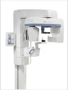
For the past 50 years, Instrumentarium Dental has been pioneering imaging systems designed for Dentists. From the original Orthopantomograph® OP1 to the most comprehensive 3-in-1 imaging system on the market today, the Orthopantomograph® OP300.
The ORTHOPANTOMOGRAPH® OP300 combines advanced panoramic imaging or Cone Beam 3D imaging giving you a truly adaptable platform.
Imaging programs:
- Adult panoramic
- Pedo panoramic
- Wide layer panoramic
- Orthogonal panoramic
- Wide arch panoramic
- Bitewing imaging
- TMJ imaging
- Cone beam imaging (4cmx6cm, 8cmx6cm)
Imaging programs:
- MULTILAYER PANORAMIC: Increases thickness of focal area compared to traditional panoramic imaging providing 5 panoramic layers in one scan.
- SMARTVIEW™ SCOUT IMAGE: A two-dimensional scout image is taken before the 3D exam, allowing for volume adjustment to ensure perfect patient positioning.
- PROVEN 5-POINT POSITIONING SYSTEM: Ensures easy patient positioning while eliminating unwanted movement during exposure
3D features:
- 6 cm x 4cm – A small FOV optimized for local diagnostics like single implant planning, 3rd molar extractions, and endodontic procedures, keep the patient dose at a substantially reduced level.
- 6 cm x 8 cm – FOV covering complete dental arch for multiple implant placement and operations using surgical guides.
Selectable 3D resolution:
- Standard resolution – Fast Reconstruction, low dose
- High resolution – For more detailed diagnostics
Potential applications:
- Panoramic X-rays
- 3D Imaging for Computer Guided Implant Placement
- Impacted or Over retained Wisdom Teeth Imaging,